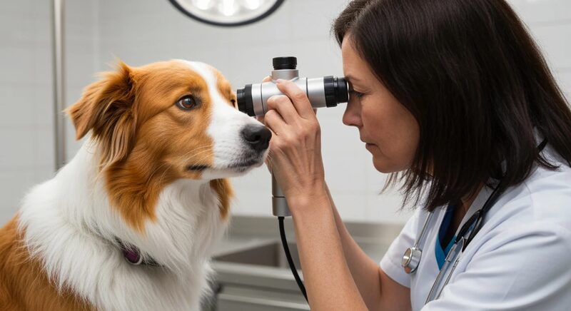
Collie Eye Anomaly: A Genetic Condition Written Into the Herding Breed Blueprint
Among the inherited eye conditions affecting herding dogs, Collie Eye Anomaly occupies a unique position. It is not a disease of adult onset or gradual degeneration — it is a developmental anomaly, present from birth, in which the eye fails to form correctly during embryonic development. Yet despite being congenital, the condition spans a remarkable range of severity: from mild choroidal changes that never affect vision, to retinal detachment and blindness diagnosed in young puppies.
Understanding Collie Eye Anomaly (CEA) means engaging with a single, well-characterized genetic variant and tracing its consequences through the developmental biology of the eye. For breeders working with Rough Collies, Smooth Collies, Border Collies, Shetland Sheepdogs, and Australian Shepherds, CEA represents one of the most prevalent genetic health issues in the gene pool — and one of the most tractable, because testing is accurate and widely available.
The Genetic Basis: A Deletion in NHEJ1
The causative mutation for CEA was identified in 2007 through a landmark study that examined the choroidal hypoplasia phenotype across multiple herding breeds. The mutation is a deletion of approximately 7.8 kilobases on canine chromosome 37, affecting the NHEJ1 gene — formally named Non-Homologous End Joining Factor 1.
This deletion disrupts a gene involved in DNA repair, but its relevance in CEA is specifically about its role in choroidal development. The choroid is the vascular layer situated between the retina and the outer sclera of the eye. During embryonic development, NHEJ1 appears to influence the proliferation and organization of cells that form the choroid. When both copies of the functional gene are absent — that is, when a dog is homozygous for the deletion — choroidal development is disrupted, and the characteristic changes of CEA result.
CEA is inherited as an autosomal recessive condition in its classical form, meaning a dog must inherit two copies of the deletion (one from each parent) to express the disease. Dogs carrying only one copy are unaffected and phenotypically indistinguishable from clear dogs without testing.
The Spectrum of Clinical Severity
What makes CEA clinically interesting — and sometimes challenging for both breeders and veterinarians — is that the severity of the condition is highly variable, even among dogs with identical genotypes.
Choroidal Hypoplasia
The mildest and most common form is choroidal hypoplasia, in which patches of the choroid are underdeveloped, appearing as pale, thinned areas when examined with an ophthalmoscope. These changes are located lateral to the optic disc and represent the primary lesion in most CEA-affected dogs. In its mild form, choroidal hypoplasia does not impair vision. The dog sees normally and may live its entire life without any clinical consequences attributable to the condition.
Breed health surveys consistently find that the majority of genetically affected dogs in Collie populations have only choroidal hypoplasia, often of minimal extent. This high frequency of subclinical disease is one reason CEA has persisted at such elevated frequency despite decades of selective breeding: many affected dogs showed no detectable abnormality under casual examination.
Coloboma
A more significant manifestation is coloboma, a term referring to a defect in the optic disc or surrounding structures caused by incomplete closure of the embryonic optic fissure. In CEA, colobomas occur at or adjacent to the optic disc. Small optic disc colobomas may produce only minor visual field distortions, but larger defects increase the risk of subsequent retinal detachment. When a coloboma is identified in a CEA-affected dog, it serves as an indicator for more intensive monitoring.
Retinal Detachment and Hemorrhage
The most severe manifestation of CEA is retinal detachment, which can be focal or complete. When detachment occurs, the photoreceptors lose their contact with the underlying retinal pigment epithelium and choroid, and degeneration follows. A dog with complete retinal detachment in both eyes is functionally blind.
Retinal detachment in CEA is not inevitable even in severely affected dogs, but its risk is highest in dogs with the most extensive choroidal changes and colobomas. Hemorrhage within the eye can accompany detachment or, in some cases, precede it. Unlike the progressive retinal atrophies, detachment in CEA typically occurs in young dogs — often within the first year of life in severe cases — rather than in middle or old age.
Why So Many Herding Breeds?
The prevalence of CEA across multiple herding breeds reflects the common ancestry of these populations before breed separation. Genetic studies using microsatellite markers and more recently whole-genome approaches have established that the NHEJ1 deletion appears to have originated in a single ancestral herding dog population, before the lineages that would become Rough Collies, Smooth Collies, Border Collies, Shetland Sheepdogs, and Australian Shepherds diverged into separate breeds.
This origin story has an important implication: the mutation reached extremely high frequencies in all these breeds before organized breeding registries existed or before breeders would have had any mechanism to systematically select against it. By the time the condition was characterized and testing became available, the deletion was present in a large fraction of every affected breed population.
Published estimates of carrier and affected dog frequencies are sobering. In Rough and Smooth Collies, the breeds where CEA was first described and most thoroughly studied, surveys from the late twentieth century found that affected dogs (homozygous for the deletion) constituted 70-90% of some population samples. The introduction of DNA testing has allowed more systematic breeding decisions, but allele frequency reduction across an entire breed population is a generational process.
This high allele frequency is directly connected to the population-level consequences of genetic bottlenecks that shaped these breeds. When populations pass through periods of severe size restriction, deleterious recessive alleles can drift to high frequency regardless of any selection pressure against them.
The Complication of “Go Normal” Dogs
CEA presents a well-documented diagnostic peculiarity that every herding breed breeder and veterinarian should understand: the phenomenon of “go normal” dogs.
In the weeks after birth, the retina is still developing and pigmenting. In some mildly affected puppies, the pale areas of choroidal hypoplasia that are clearly visible during ophthalmoscopic examination at three to six weeks of age become progressively obscured by normal retinal pigmentation as the puppy matures. By the time the dog reaches adulthood, the choroidal changes may be entirely undetectable on clinical examination.
This means that a dog examined as an adult might receive a clinical “clear” result, despite being genetically affected and capable of passing the NHEJ1 deletion to offspring. This is precisely the category of CEA-affected dog that perpetuated the condition through generations of Collie breeding — they appeared normal to experienced breeders performing ophthalmologic examination, were bred freely, and maintained the deletion in the population.
The existence of go-normal dogs is the primary reason that DNA testing has transformed CEA management, and why clinical eye examination alone is insufficient for making breeding decisions. A DNA test that identifies the NHEJ1 deletion reports a dog’s genotype accurately regardless of its age or the degree to which retinal pigmentation obscures the choroidal changes. For a comprehensive overview of how to select and interpret canine genetic tests, including sample collection and laboratory choice, see our guide to canine DNA testing.
Ophthalmologic Examination Still Has a Role
Even though DNA testing is the cornerstone of CEA management, clinical ophthalmologic examination by a board-certified veterinary ophthalmologist remains valuable and should not be abandoned in favor of genetic testing alone.
First, the DNA test detects only the known NHEJ1 deletion. Atypical or modifier variants affecting CEA expression may not be captured, and other ocular abnormalities — including primary lens luxation, persistent pupillary membranes, and other developmental anomalies — require clinical detection.
Second, for dogs that will be shown or certified through registry health schemes, ophthalmologic certification through systems such as CAER (Canine Eye Registry Foundation) in North America or BVA/KC scheme in the UK provides a standardized clinical record that complements molecular testing. These examinations should ideally be performed at the recommended age window — before retinal pigmentation becomes complete — to maximize the sensitivity for detecting mild choroidal changes in dogs where DNA testing may not be available.
Third, severity assessment in genetically affected dogs requires ophthalmologic examination. The DNA test identifies a dog as homozygous affected but cannot predict whether the dog has minimal choroidal hypoplasia or more serious colobomas and risk of detachment. Clinical grading of severity informs the prognosis for individual dogs and guides monitoring decisions.
Breeding Strategy: Managing a High-Frequency Allele
The challenge CEA poses for herding breed breeders differs from the management problem posed by rarer recessive conditions. When allele frequencies are as high as they are in Collies and Shelties, a strategy of avoiding all carriers entirely would effectively eliminate most of the breed population and create a profound loss of genetic diversity that would almost certainly cause more harm than the disease itself.
The practical framework used by most breed health organizations acknowledges this reality:
Clear dogs (no copies of the deletion) should be paired together whenever possible and are the preferred option for maintaining clear offspring.
Carrier × clear pairings are acceptable and produce no affected offspring while maintaining the carrier dog’s contribution to genetic diversity. Fifty percent of offspring will be carriers, but none will be affected.
Affected × clear pairings are strongly discouraged because all offspring will be carriers, and there is no breeding context in which a homozygous affected dog cannot be replaced by a carrier animal.
Affected × carrier or affected × affected pairings should not occur under any circumstances.
The goal for breed populations is gradual reduction of the deletion allele frequency over multiple generations through the consistent application of carrier × clear pairings and the preferential selection of clear breeding stock wherever this can be done without further narrowing genetic diversity. Progress has been measurable in breeds where testing adoption rates have been high. Breeders who test consistently and make results available through public registries contribute to population-level progress, not just the health of their individual litters.
Implications for Puppy Buyers
For prospective owners of Rough Collies, Smooth Collies, Shetland Sheepdogs, Border Collies, or Australian Shepherds, CEA testing of breeding stock is a reasonable expectation to hold. A responsible breeder should be able to provide DNA testing documentation for both parents and should be transparent about whether any breeding stock is a carrier or affected.
A puppy from two DNA-clear parents cannot be affected. A puppy from a carrier × clear pairing has a 50% chance of being a carrier but cannot be affected. Understanding these probabilities allows puppy buyers to make informed decisions and to prioritize breeders who demonstrate commitment to genetic health management.
Puppy buyers outside North America and the UK are working with a different certification system: continental Europe’s ECVO ophthalmologist network runs its own eye scheme, examined alongside, not instead of, the NHEJ1 DNA result. Dutch-speaking buyers researching a Collie litter can read how the two are meant to work together on Colliewijzer’s page on CEA and the ECVO eye exam, including why breeders test puppies before they leave the litter.
CEA is ultimately a manageable condition. The severe forms — retinal detachment and blindness — represent a small proportion of affected dogs. The availability of accurate DNA testing, combined with the growing culture of transparency in herding breed health testing, has created the conditions for meaningful, generational reduction of this condition across the breeds where it has historically been most prevalent.
Frequently Asked Questions
Which herding breeds are most affected by Collie Eye Anomaly?
Rough and Smooth Collies historically show the highest carrier and affected frequencies, with surveys from earlier decades finding affected dogs (homozygous for the NHEJ1 deletion) in 70 to 90 percent of some population samples. Shetland Sheepdogs, Border Collies, and Australian Shepherds are also significantly affected. All share common herding dog ancestry that predates the establishment of separate breed registries.
Can a CEA-affected dog have normal vision?
Yes. The majority of genetically affected dogs have only mild choroidal hypoplasia, which does not impair vision. Only dogs with more severe manifestations — colobomas, retinal detachment, or hemorrhage — experience visual impairment. A dog certified as genetically affected by DNA testing may still have entirely normal functional vision throughout its life.
What are “go normal” dogs and why do they matter?
Go-normal dogs are CEA-affected dogs whose mild choroidal changes become obscured by normal retinal pigmentation as the puppy matures. What was visible at three to six weeks of age becomes undetectable on adult ophthalmoscopic examination. These dogs test genetically positive but appear clinically clear as adults, which historically allowed the NHEJ1 deletion to persist through generations of apparently normal breeding stock. DNA testing eliminates this diagnostic blind spot.
Is CEA the same as progressive retinal atrophy?
No. CEA is a developmental condition present from birth, caused by the NHEJ1 deletion and affecting choroidal development. Progressive retinal atrophy (PRA) is a family of degenerative conditions in which photoreceptors progressively die in mature dogs. The two conditions can coexist in the same dog, which is why both conditions should be tested in breeds susceptible to each. Our article on progressive retinal atrophy in herding breeds provides detailed coverage of PRA genetics.
Should I buy a puppy whose parents are CEA carriers?
A puppy from a carrier × clear pairing cannot be affected — only carriers or clear dogs result from this cross. If the breeder has DNA tested both parents, a puppy from this pairing carries no risk of CEA-related visual impairment. Many responsible breeders use high-quality carrier dogs in their programs when those dogs offer valuable genetic diversity or other important health and working qualities, and this is a defensible choice as long as all offspring are tested before any future breeding decisions are made.
Collie Eye Anomaly follows a simple autosomal recessive pattern, which is why a single DNA test can classify a dog as clear, carrier, or affected with near certainty. Hip dysplasia does not behave that way. Its inheritance is polygenic, shaped by many loci of small effect plus environmental factors such as growth rate and exercise during puppyhood, so no genotype test can predict an individual dog’s outcome. Breeders managing both conditions in the same litter therefore rely on DNA results for CEA and on joint screening in herding breeds for hip status, combining the two when selecting breeding pairs.